Is VBT the Right Question for Your Family?
If you are reading this, you have probably already heard the word "fusion" in a clinic and felt your stomach drop. Most parents do. Spinal fusion is a reliable operation, and for many teenagers it remains the right one. But for a specific group of younger patients there is now a second door: vertebral body tethering, usually shortened to VBT.
VBT is for a child whose spine is still growing, whose curve has passed the point where a brace can hold it, and whose curve still bends easily on X-ray. Get those three things right and the tether can work with growth rather than against it. Get any of them wrong and it is the wrong operation. So before anything else, here is the honest checklist.
| Curve size at surgery | Roughly 35° to 70° (Cobb angle). Below that, bracing and observation are still on the table; above it, the tether cannot do enough. |
|---|---|
| Skeletal maturity | The spine must still be immature: typically Risser 0 to 2, with the hand X-ray (Sanders stage) confirming growth ahead. Growth is the engine of the correction. |
| Flexibility | The curve should correct substantially on a side-bending X-ray. A stiff curve does not respond to a tether. |
| Approach | Endoscopic (thoracoscopic), minimally invasive. Three to five small incisions on the side of the chest instead of one long incision down the back. |
| What is implanted | One titanium screw in each vertebra across the curve, joined by a flexible polymer cord on the convex (outer) side. No rods, no bone graft, no fusion. |
| How it corrects | Partly on the table when the cord is tensioned, then progressively over one to three years as growth on the tethered side slows and the concave side catches up. |
| Movement afterwards | The tethered segments keep bending and twisting. That is the whole point of choosing VBT. |
| Main trade-off | Higher chance of needing a second procedure than fusion. Cord breakage and over-correction are the two things we watch for. |
If your child has not yet been diagnosed or you are still at the "what is scoliosis" stage, start with our overview of Adolescent Idiopathic Scoliosis and come back here.
See the Operation in Three Dimensions
Words only go so far with an operation like this, so I built a model. It opens on a growing spine with a 50° curve, walks through the operation step by step, then lets you set the curve size and years of growth remaining yourself to see how the answer changes. Drag to rotate, scroll to zoom. On a phone, turn it sideways.
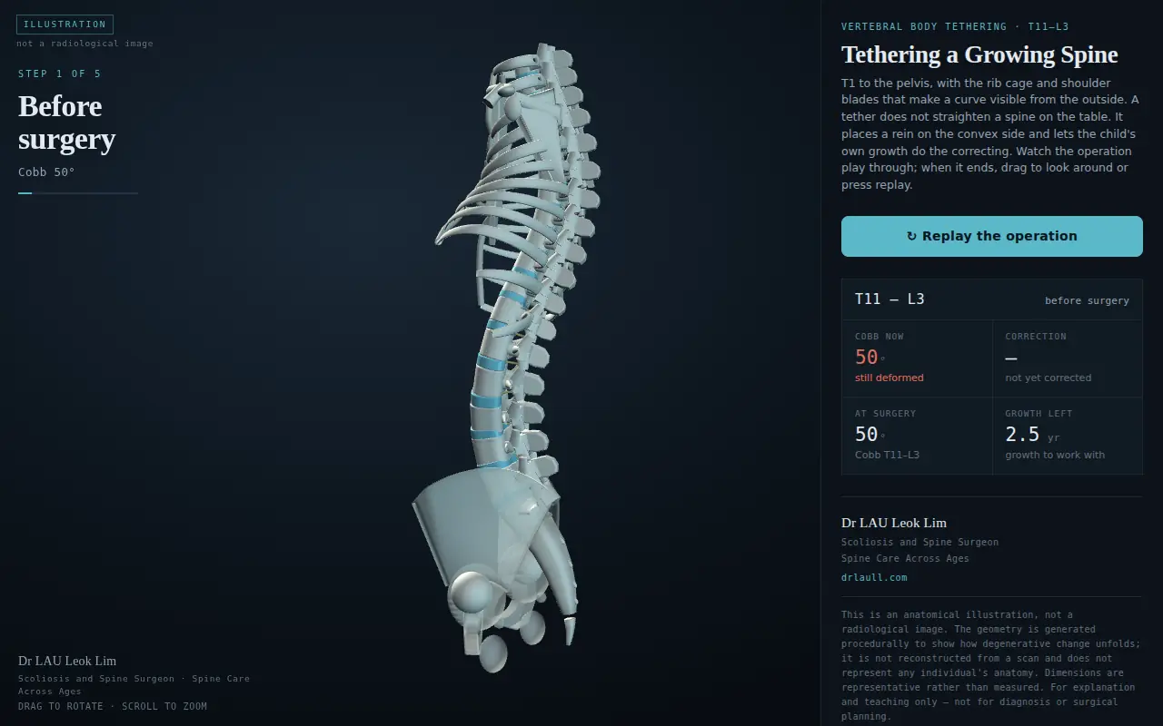
Loading the interactive model…
Open the model full-screen · Press ↻ Replay the operation inside the model to watch it again.
Educational illustration only. The anatomy is generated, not reconstructed from any patient's scan, and the correction shown is a simplified model of growth modulation. It does not predict any individual's result and does not replace clinical assessment or X-rays.
How a Tether Straightens a Spine Without Fusing It
Every growing bone has growth plates, and vertebrae are no different. A scoliotic curve sets up a vicious cycle: the vertebrae on the inside of the curve are squeezed, so their growth plates slow down, while the outside of the curve is stretched and grows faster. The faster the outside grows, the bigger the curve, the more the inside is squeezed. Left alone in a growing child, the curve feeds itself.
VBT turns that same biology around. The cord is anchored to the fast-growing convex side and tensioned. The pressure it applies gently slows growth on that side. The concave side, now relieved of some of its load, keeps growing at its normal pace. Over the next growth spurt the two sides come back into balance and the curve unwinds. Surgeons call this growth modulation. I describe it to families as re-balancing a growth mismatch, because that is exactly what it is.
What happens on the day
The operation is done through the side of the chest using a camera and long instruments, the same thoracoscopic technique used for many chest procedures. There is no long incision down the back and no stripping of the back muscles. Three to five small cuts are placed between the ribs on the convex side. Under X-ray guidance a screw is placed into the side of each vertebra across the curve, one level at a time. The cord is threaded through the screw heads from top to bottom, then tensioned segment by segment. The curve visibly reduces on the table, usually by about half. Most children are walking the next day and home within three to five days.
What happens over the next two years
This is the part that feels strange to parents: after surgery, the correction keeps improving without anyone doing anything. We take standing X-rays at regular intervals to watch it. The aim is a curve that settles somewhere in the teens or low twenties in degrees, in a spine that still bends. Because the correction depends on growth, it is not finished until growth is, which is why follow-up runs until the spine is mature.
Who Is a Candidate, and Who Is Not
Candidacy is where most of the judgement in VBT lives, and it is where I spend most of the first consultation. The three questions are the same for every child.
1. Is the curve in the surgical range?
Between roughly 35° and 70°. A curve that has reached 35° to 40° in a growing child has, in most cases, shown that a brace is not going to hold it. Above 70° the curve is usually too large and too stiff for a tether to correct adequately, and fusion becomes the safer recommendation. If bracing has already been tried and the curve progressed through it, that history is relevant; our section on bracing in AIS explains what a brace can and cannot do.
2. Is there enough growth left?
This is assessed from age, menarche in girls, the Risser sign on the pelvis X-ray and the Sanders stage on a hand X-ray. The ideal candidate is at Risser 0 to 2 and Sanders 2 to 4, with a meaningful growth spurt still to come. Too little growth and the tether gives only the on-table correction. Too much growth in a young child, and the curve can carry on past straight into the opposite direction, which is called over-correction.
3. Is the curve flexible?
A side-bending X-ray tells us how much of the curve is structural and how much is still supple. A curve that reduces well on bending is a curve that a tether can work with.
VBT or Fusion: A Fair Comparison
Parents often arrive wanting VBT because it sounds gentler, and it is a smaller operation on the day. But "smaller on the day" is only part of the picture. Here is how the two compare across the things families actually ask about.
| Spinal movement | VBT: tethered levels keep moving. Fusion: fused levels become one solid block; levels above and below take over. |
|---|---|
| Incision and recovery | VBT: keyhole, less blood loss, shorter operation, usually home in 3 to 5 days. Fusion: open, longer operation, usually home in 4 to 6 days. |
| How much correction | VBT: about half on the table, more with growth; final result depends on growth. Fusion: most of the correction on the table, predictable and permanent. |
| Predictability | VBT: less predictable, because it relies on biology. Fusion: more predictable. |
| Chance of a second operation | VBT: higher, most often for cord breakage, over-correction or a curve that keeps progressing. Fusion: lower. |
| Return to sport | VBT: typically earlier. Fusion: a structured return over several months; see our feature on competitive sport after scoliosis surgery. |
| If it does not work | VBT: fusion remains available later. Fusion: cannot be undone. |
Put simply: VBT trades some predictability for preserved motion and a smaller operation, and it only makes that trade well in the right child. The published evidence says the same. Across pooled studies, tethering corrects the main curve by roughly half, keeps the spine mobile, gets children home faster, and carries a higher revision rate than fusion. Cord breakage has been reported in somewhere between one in eight and one in four patients over the first few years, though a broken cord does not always need anything done about it if the curve has already settled. I would rather you read those numbers here than discover them later.
What to Expect: Before, During and After
Before surgery
Standing whole-spine X-rays, bending films, a hand X-ray for Sanders staging and an MRI of the whole spine to rule out anything unusual in the spinal cord. A CT scan may be used to plan screw size and position. We talk through the model above together so that both parent and teenager understand what is being asked of the spine.
The hospital stay
The operation takes two to four hours depending on the number of levels. A small chest drain is usually left in for a day or two because the surgery is performed inside the chest cavity. Most patients are up and walking the next day, and discharged when they are comfortable and the drain is out.
The first six weeks
Walking is encouraged from day one. No brace is required. Heavy lifting, contact sport and deep bending are avoided while the screws settle into bone. School can usually resume within two to three weeks.
Longer term
Gradual return to sport from about three months, with full activity once the surgical site has healed and the X-rays are stable. Standing X-rays every four to six months until the spine is mature, then yearly for a period after that. The curve should be seen to improve with each growth check. If it improves too quickly, or a cord breaks and the curve moves, that is picked up early on these films.
For the wider picture on living well with scoliosis through the teenage years, our Embrace patient guidebook and tips for living with scoliosis are written for exactly this stage.
Risks and Honest Limitations
Every spinal operation carries general risks: infection, bleeding, anaesthetic risks, and the rare but serious risk of nerve or spinal cord injury, which is monitored throughout surgery with continuous nerve signal recording. VBT adds a few of its own that families should know before choosing it.
- Cord breakage. The polymer cord can fatigue and break, usually a year or more after surgery. Often the curve has already settled and nothing needs to be done. Sometimes the curve drifts back and a revision or conversion to fusion is needed.
- Over-correction. In a child with a lot of growth left, the curve can continue past straight. This is watched for on every X-ray and can be treated by releasing or loosening the cord.
- Under-correction. If growth turns out to be less than estimated, the curve may not improve as much as hoped. Fusion remains an option later.
- Chest-related issues. Because the operation is performed through the chest, a small collection of fluid or air around the lung can occur and is managed with the drain.
- It is still a newer operation. Long-term results beyond ten years are only now being reported. Fusion has decades of follow-up behind it.
None of this is a reason to dismiss VBT. It is a reason to choose it carefully, in the right child, with eyes open.
If you are earlier in the journey and still working out whether a curve is there at all, start with the visual guide to what scoliosis looks like, which uses the same 3D model.
Cost in Singapore
VBT uses a purpose-made implant system and specialised endoscopic equipment, so implant costs are generally higher than fusion, while hospital stay is usually shorter. Fees vary with the number of levels, the hospital and your insurance or MediSave arrangements. Our separate article on the cost of scoliosis surgery in Singapore explains how the components add up and what to ask before you commit. A written estimate is provided after the consultation once the number of levels is known.
Frequently Asked Questions (FAQ)
1. Is vertebral body tethering minimally invasive?
Yes. It is performed thoracoscopically, through a few small incisions between the ribs using a camera and long instruments, rather than through a long open incision down the back. Muscle disruption and blood loss are considerably less than with open fusion.
2. What curve size is suitable for VBT?
Roughly 35° to 70° Cobb angle in a child who is still growing and whose curve is flexible on bending X-rays. Below 35° bracing is usually still appropriate; above 70° the tether cannot achieve enough correction and fusion is generally recommended.
3. My teenager is 15. Is it too late?
Not necessarily, but it depends on skeletal maturity rather than age. A 15-year-old girl who has passed menarche and is at Risser 4 is usually too mature; a 15-year-old boy who has not yet had his growth spurt may be an excellent candidate. The hand X-ray decides.
4. Does the spine straighten immediately?
Partly. Tensioning the cord corrects roughly half of the curve on the operating table. The rest of the correction happens over the following one to three years as growth is modulated. You will see it improve on each follow-up X-ray.
5. Will my child be able to bend and play sport?
Yes. Preserving movement is the main reason VBT exists. Most patients return to non-contact activity within three months and to full sport once healed and cleared. Tethered segments retain most of their flexibility.
6. What happens if the tether breaks?
Cord breakage is the most common VBT-specific complication. If the curve has already stabilised, often nothing needs to be done. If the curve drifts, options include re-tensioning, replacing the cord, or converting to fusion. This is one of the reasons follow-up X-rays continue until maturity.
7. Can VBT be done on the lumbar spine?
Yes. Thoracic curves are approached through the chest, and thoracolumbar or lumbar curves through a small incision on the flank. The model on this page shows a thoracolumbar construct from T11 to L3.
8. Is VBT approved and established?
Tethering systems have regulatory approval in the United States (through a Humanitarian Device Exemption granted in 2019) and are in use in many countries, including Singapore. It is a newer operation than fusion, and long-term data continue to accumulate.
9. Who should I see?
A spine surgeon with specific training in both paediatric deformity and thoracoscopic technique, who also performs fusion and can advise honestly on which operation fits your child. Bring all previous X-rays to the first consultation.
Selected Evidence
The comparisons above draw on peer-reviewed pooled analyses, listed here for families and colleagues who would like to read further.
- Al-Naseem AO, et al. Posterior spinal fusion versus vertebral body tethering for paediatric scoliosis: a meta-analysis of comparative studies. Spine Deformity. 2025;13(3):681-694. doi:10.1007/s43390-025-01050-x
- Abdelaal M, et al. Anterior vertebral body tethering versus posterior spinal fusion in adolescent idiopathic scoliosis: a systematic review and meta-analysis of comparative outcomes. Journal of Clinical Medicine. 2025;14(19):6707. doi:10.3390/jcm14196707
- Hevia E, et al. Emerging growth modulation techniques in treatment of adolescent idiopathic scoliosis: a meta-analysis of the outcomes and safety of anterior vertebral body tethering and ApiFix. Asian Spine Journal. 2026. doi:10.31616/asj.2025.0612